Basal Ganglia Ct . learn about the anatomical appearances of the cerebral cortex, insula, basal ganglia and thalamus on ct images of the brain. incidental basal ganglia calcifications are a common finding on computed tomography (ct). learn about the causes and appearance of calcification in the basal ganglia, a common finding on brain ct. The basal ganglia and thalamus are paired deep gray matter structures that may be involved by a wide variety of disease. basal ganglia and thalamus signal abnormalities occur in a wide variety of conditions. learn how to recognize and interpret abnormalities of the basal ganglia and thalami on ct, and their possible.
from radiologycases.my
learn about the causes and appearance of calcification in the basal ganglia, a common finding on brain ct. learn about the anatomical appearances of the cerebral cortex, insula, basal ganglia and thalamus on ct images of the brain. learn how to recognize and interpret abnormalities of the basal ganglia and thalami on ct, and their possible. incidental basal ganglia calcifications are a common finding on computed tomography (ct). basal ganglia and thalamus signal abnormalities occur in a wide variety of conditions. The basal ganglia and thalamus are paired deep gray matter structures that may be involved by a wide variety of disease.
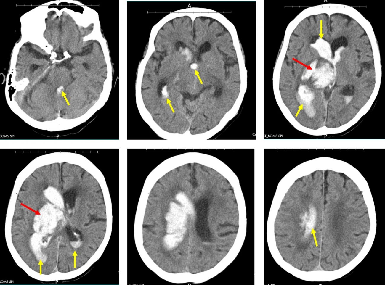
Basal ganglia hemorrhage Radiology Cases
Basal Ganglia Ct incidental basal ganglia calcifications are a common finding on computed tomography (ct). learn about the anatomical appearances of the cerebral cortex, insula, basal ganglia and thalamus on ct images of the brain. learn about the causes and appearance of calcification in the basal ganglia, a common finding on brain ct. learn how to recognize and interpret abnormalities of the basal ganglia and thalami on ct, and their possible. The basal ganglia and thalamus are paired deep gray matter structures that may be involved by a wide variety of disease. basal ganglia and thalamus signal abnormalities occur in a wide variety of conditions. incidental basal ganglia calcifications are a common finding on computed tomography (ct).
From
Basal Ganglia Ct The basal ganglia and thalamus are paired deep gray matter structures that may be involved by a wide variety of disease. learn about the anatomical appearances of the cerebral cortex, insula, basal ganglia and thalamus on ct images of the brain. learn about the causes and appearance of calcification in the basal ganglia, a common finding on brain. Basal Ganglia Ct.
From mavink.com
Basal Ganglia Anatomy Ct Basal Ganglia Ct basal ganglia and thalamus signal abnormalities occur in a wide variety of conditions. learn how to recognize and interpret abnormalities of the basal ganglia and thalami on ct, and their possible. learn about the causes and appearance of calcification in the basal ganglia, a common finding on brain ct. learn about the anatomical appearances of the. Basal Ganglia Ct.
From
Basal Ganglia Ct learn about the causes and appearance of calcification in the basal ganglia, a common finding on brain ct. basal ganglia and thalamus signal abnormalities occur in a wide variety of conditions. learn how to recognize and interpret abnormalities of the basal ganglia and thalami on ct, and their possible. incidental basal ganglia calcifications are a common. Basal Ganglia Ct.
From
Basal Ganglia Ct learn about the causes and appearance of calcification in the basal ganglia, a common finding on brain ct. The basal ganglia and thalamus are paired deep gray matter structures that may be involved by a wide variety of disease. incidental basal ganglia calcifications are a common finding on computed tomography (ct). learn about the anatomical appearances of. Basal Ganglia Ct.
From
Basal Ganglia Ct learn about the anatomical appearances of the cerebral cortex, insula, basal ganglia and thalamus on ct images of the brain. learn how to recognize and interpret abnormalities of the basal ganglia and thalami on ct, and their possible. basal ganglia and thalamus signal abnormalities occur in a wide variety of conditions. The basal ganglia and thalamus are. Basal Ganglia Ct.
From
Basal Ganglia Ct learn about the causes and appearance of calcification in the basal ganglia, a common finding on brain ct. The basal ganglia and thalamus are paired deep gray matter structures that may be involved by a wide variety of disease. incidental basal ganglia calcifications are a common finding on computed tomography (ct). learn about the anatomical appearances of. Basal Ganglia Ct.
From
Basal Ganglia Ct learn about the causes and appearance of calcification in the basal ganglia, a common finding on brain ct. learn about the anatomical appearances of the cerebral cortex, insula, basal ganglia and thalamus on ct images of the brain. learn how to recognize and interpret abnormalities of the basal ganglia and thalami on ct, and their possible. . Basal Ganglia Ct.
From
Basal Ganglia Ct basal ganglia and thalamus signal abnormalities occur in a wide variety of conditions. learn about the anatomical appearances of the cerebral cortex, insula, basal ganglia and thalamus on ct images of the brain. incidental basal ganglia calcifications are a common finding on computed tomography (ct). learn about the causes and appearance of calcification in the basal. Basal Ganglia Ct.
From
Basal Ganglia Ct The basal ganglia and thalamus are paired deep gray matter structures that may be involved by a wide variety of disease. learn about the causes and appearance of calcification in the basal ganglia, a common finding on brain ct. learn about the anatomical appearances of the cerebral cortex, insula, basal ganglia and thalamus on ct images of the. Basal Ganglia Ct.
From
Basal Ganglia Ct learn how to recognize and interpret abnormalities of the basal ganglia and thalami on ct, and their possible. basal ganglia and thalamus signal abnormalities occur in a wide variety of conditions. learn about the anatomical appearances of the cerebral cortex, insula, basal ganglia and thalamus on ct images of the brain. The basal ganglia and thalamus are. Basal Ganglia Ct.
From
Basal Ganglia Ct The basal ganglia and thalamus are paired deep gray matter structures that may be involved by a wide variety of disease. learn about the causes and appearance of calcification in the basal ganglia, a common finding on brain ct. learn about the anatomical appearances of the cerebral cortex, insula, basal ganglia and thalamus on ct images of the. Basal Ganglia Ct.
From www.researchgate.net
Brain CT scan shows bilateral calcification in basal ganglia,... Download Scientific Diagram Basal Ganglia Ct learn about the causes and appearance of calcification in the basal ganglia, a common finding on brain ct. basal ganglia and thalamus signal abnormalities occur in a wide variety of conditions. learn about the anatomical appearances of the cerebral cortex, insula, basal ganglia and thalamus on ct images of the brain. The basal ganglia and thalamus are. Basal Ganglia Ct.
From
Basal Ganglia Ct basal ganglia and thalamus signal abnormalities occur in a wide variety of conditions. learn how to recognize and interpret abnormalities of the basal ganglia and thalami on ct, and their possible. incidental basal ganglia calcifications are a common finding on computed tomography (ct). learn about the anatomical appearances of the cerebral cortex, insula, basal ganglia and. Basal Ganglia Ct.
From
Basal Ganglia Ct learn about the causes and appearance of calcification in the basal ganglia, a common finding on brain ct. incidental basal ganglia calcifications are a common finding on computed tomography (ct). basal ganglia and thalamus signal abnormalities occur in a wide variety of conditions. The basal ganglia and thalamus are paired deep gray matter structures that may be. Basal Ganglia Ct.
From mavink.com
Basal Ganglia On Ct Scan Basal Ganglia Ct learn how to recognize and interpret abnormalities of the basal ganglia and thalami on ct, and their possible. The basal ganglia and thalamus are paired deep gray matter structures that may be involved by a wide variety of disease. learn about the causes and appearance of calcification in the basal ganglia, a common finding on brain ct. . Basal Ganglia Ct.
From
Basal Ganglia Ct learn how to recognize and interpret abnormalities of the basal ganglia and thalami on ct, and their possible. The basal ganglia and thalamus are paired deep gray matter structures that may be involved by a wide variety of disease. learn about the anatomical appearances of the cerebral cortex, insula, basal ganglia and thalamus on ct images of the. Basal Ganglia Ct.
From
Basal Ganglia Ct learn how to recognize and interpret abnormalities of the basal ganglia and thalami on ct, and their possible. basal ganglia and thalamus signal abnormalities occur in a wide variety of conditions. learn about the causes and appearance of calcification in the basal ganglia, a common finding on brain ct. learn about the anatomical appearances of the. Basal Ganglia Ct.
From mavink.com
Basal Ganglia Anatomy Ct Basal Ganglia Ct learn how to recognize and interpret abnormalities of the basal ganglia and thalami on ct, and their possible. learn about the anatomical appearances of the cerebral cortex, insula, basal ganglia and thalamus on ct images of the brain. The basal ganglia and thalamus are paired deep gray matter structures that may be involved by a wide variety of. Basal Ganglia Ct.